
The process of visually examining the urinary bladder is an important medical procedure used to diagnose and monitor various conditions affecting the bladder. This procedure, known as cystoscopy, involves the use of a thin, flexible tube with a camera at its tip, called a cystoscope.
During a cystoscopy, the patient is typically placed under anesthesia to ensure their comfort throughout the procedure. The cystoscope is then gently inserted into the urethra and guided into the bladder. The camera at the tip of the cystoscope allows the urologist to visually inspect the bladder walls and surrounding structures in real-time.
This procedure allows healthcare professionals to identify and evaluate various conditions that may be affecting the urinary bladder. These conditions can include the presence of tumors or polyps, inflammation or infection of the bladder lining, and structural abnormalities of the bladder or urethra. In addition to visualization, biopsies can be obtained during a cystoscopy to further evaluate any suspicious areas or lesions.
Overall, the process of visually examining the urinary bladder through cystoscopy provides valuable insight into the health and function of this crucial organ. By identifying and diagnosing any abnormalities or conditions, healthcare professionals can then develop an appropriate treatment plan to address the patient’s specific needs and promote their overall well-being.
What is the urinary bladder?
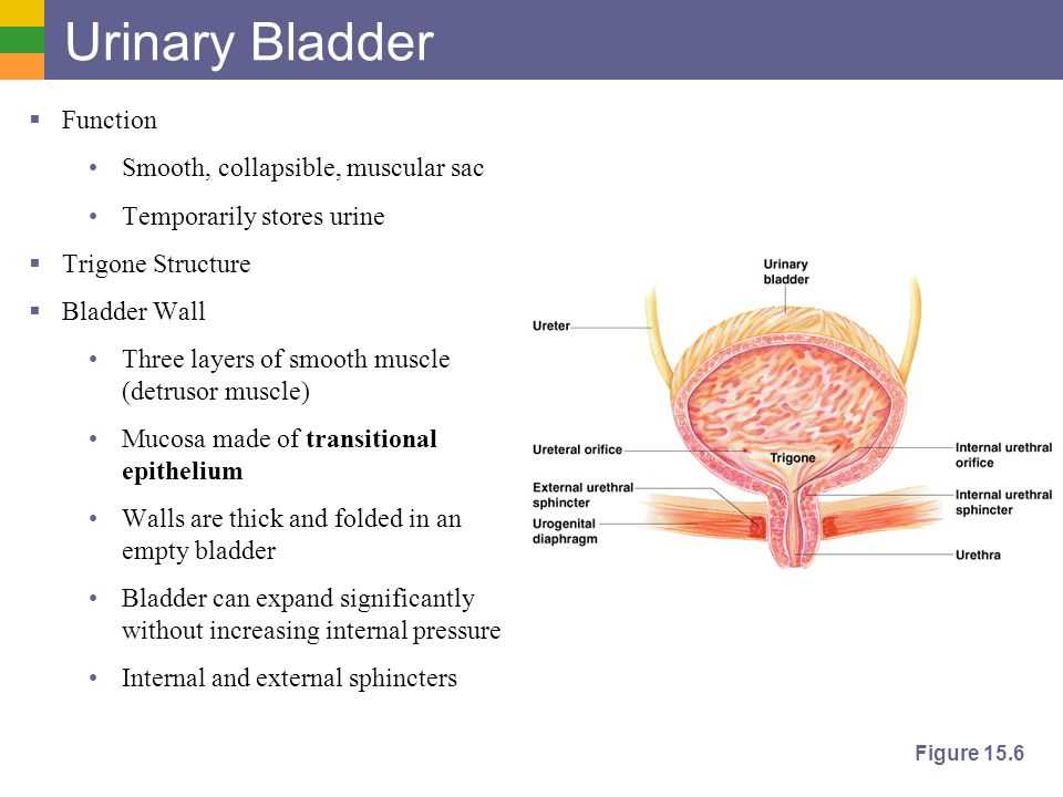
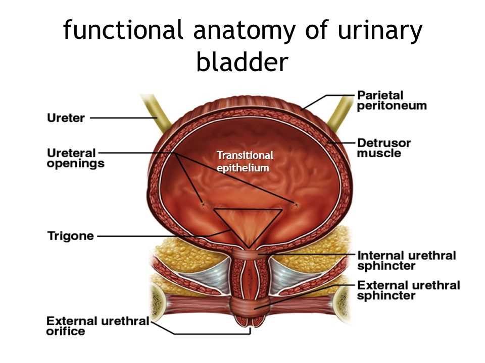
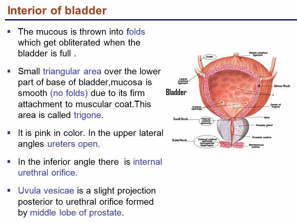
The urinary bladder is a hollow organ located in the pelvis, which plays a vital role in the urinary system. It is a muscular sac that stores urine produced by the kidneys before it is eliminated from the body through the urethra. The bladder is lined with a specialized layer of cells called urothelium, which allows for efficient storage of urine without leakage.
The structure of the bladder consists of three layers of muscle: the inner layer of smooth muscle called the detrusor muscle, the middle layer of connective tissue, and the outer layer of smooth muscle. These muscles work together to contract and expand, allowing the bladder to store increasing amounts of urine as it fills and then contract to expel the urine during urination.
Key Functions of the Urinary Bladder:
- Storage: The main function of the bladder is to store urine until it can be expelled from the body. The muscular walls of the bladder can stretch and accommodate increasing volumes of urine without causing discomfort or leakage.
- Control: The bladder has a complex system of nerves and muscles that help regulate the timing and control of urination. This allows individuals to voluntarily choose when to empty their bladders.
- Protection: The bladder helps protect the urinary system by preventing the backflow of urine into the kidneys. When the bladder is full, a valve called the urethral sphincter closes, preventing urine from traveling back up the ureters.
In summary, the urinary bladder is a vital organ involved in the storage and elimination of urine. Its muscular walls, specialized lining, and control mechanisms allow for efficient urine storage and controlled urination.
Importance of visually examining the urinary bladder
The visual examination of the urinary bladder, also known as cystoscopy, plays a crucial role in diagnosing and evaluating various urological conditions. This procedure allows healthcare professionals to directly observe the inner lining of the bladder and detect any abnormalities or potential issues that may require further investigation or treatment.
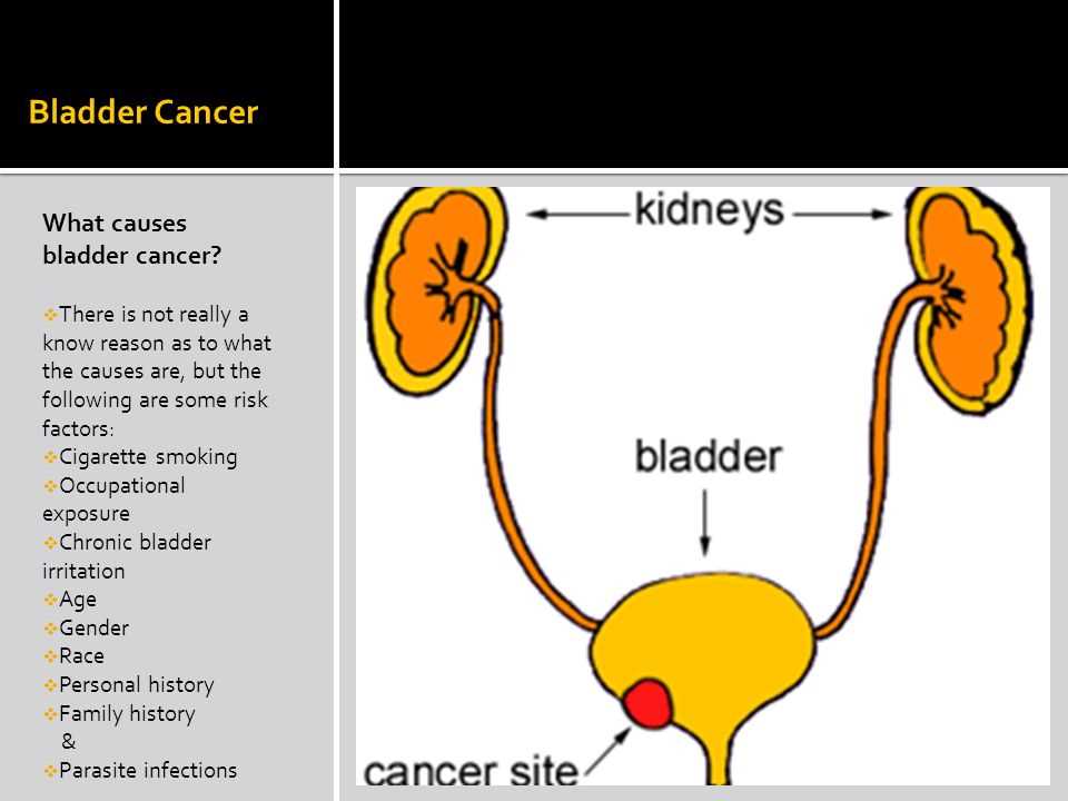
One of the primary reasons for visually examining the urinary bladder is to diagnose and monitor bladder tumors. Cystoscopy enables doctors to identify and evaluate the size, location, and characteristics of any tumors or suspicious areas within the bladder. This information is vital in determining the appropriate treatment approach, such as surgical removal or targeted therapies, and in monitoring the response to treatment.
In addition to detecting tumors, cystoscopy is also essential in investigating hematuria, which is the presence of blood in the urine. By directly visualizing the bladder, healthcare professionals can identify the source of bleeding, whether it’s due to bladder stones, urinary tract infections, kidney stones, or other underlying conditions. This allows for timely and accurate diagnosis, as well as appropriate management of the underlying cause.
- Cystoscopy is also used for the following purposes:
- – Evaluating and managing urinary incontinence
- – Diagnosing and treating recurrent urinary tract infections
- – Assessing the effectiveness of previous bladder surgeries or treatments
- – Identifying bladder stones or foreign bodies
- – Guiding the placement of urinary catheters or other devices
In summary, the visual examination of the urinary bladder through cystoscopy is a critical tool in the diagnosis, treatment, and monitoring of various urological conditions. By directly observing the bladder, healthcare professionals can accurately detect and evaluate tumors, investigate the source of hematuria, and assess the effectiveness of previous treatments. This procedure not only helps in improving patient outcomes but also guides the development of appropriate treatment plans and interventions.
Preparation for the examination
Before undergoing a visual examination of the urinary bladder, there are several steps that need to be taken to ensure a successful and accurate procedure. These preparations involve both the patient and the medical staff involved in the examination.
Patient preparation:
- The patient should inform the medical staff about any allergies or medical conditions they may have, as this information can affect the choice of contrast agents or medications used during the examination.
- The patient may be asked to fast for a certain period of time before the examination, especially if a contrast agent will be used. This is typically done to ensure clear visualization of the bladder and surrounding structures.
- The patient may need to provide a urine sample prior to the examination to check for any signs of infection or other abnormalities.
- If necessary, the patient may need to undergo a bowel preparation to ensure that the intestines are empty and do not interfere with the examination.
Medical staff preparation:
- The medical staff should review the patient’s medical history and any relevant previous examination results to better understand the context of the examination.
- The medical staff should ensure that all necessary equipment and supplies are properly sterilized and prepared for the examination.
- The medical staff should explain the procedure to the patient, including any potential risks or discomfort that may be associated with the examination.
- If a contrast agent will be used, the medical staff should ensure that it is properly prepared and available for use during the examination.
- The medical staff should also ensure that the imaging equipment is properly calibrated and functioning correctly before the examination begins.
Obtaining a medical history
The process of visually examining the urinary bladder begins with obtaining a comprehensive medical history of the patient. This step is crucial in understanding the patient’s background, medical conditions, and any special considerations that need to be taken into account during the examination.
To obtain a medical history, the healthcare provider typically starts by asking the patient a series of questions. These questions may include inquiries about the patient’s current symptoms, such as pain or discomfort in the urinary tract, frequent urination, or blood in the urine. The provider may also ask about the patient’s history of urinary tract infections, kidney stones, or any previous surgeries related to the bladder or urinary system.
In addition to specific bladder-related questions, the healthcare provider will also ask about the patient’s overall health and medical history. This may include questions about chronic conditions such as diabetes or hypertension, medication use, allergies, and any previous surgeries or medical procedures the patient has undergone. It is important for the healthcare provider to have a complete understanding of the patient’s medical history in order to ensure a safe and effective examination of the urinary bladder.
Physical examination
The physical examination is an important step in visually examining the urinary bladder. It involves the use of various tools and techniques to gather information about the bladder’s size, shape, and condition.
One common tool used in the physical examination is a cystoscope, which is a thin, flexible tube with a camera on the end. This allows the doctor to visualize the inside of the bladder and assess any abnormalities or abnormalities.
In addition to the cystoscope, the doctor may also use a resectoscope, which is a similar instrument but with a loop or wire attached. This allows for the removal or biopsy of tissue from the bladder for further examination.
During the physical examination, the doctor may also use manual palpation techniques to assess the bladder. This involves gently pressing on the abdomen to feel for any abnormalities or changes in the bladder’s shape or size.
Overall, the physical examination is an essential part of visually examining the urinary bladder. It allows the doctor to gather important information about the bladder’s condition and helps guide further diagnostic and treatment decisions.
Laboratory Tests
The process of visually examining the urinary bladder involves various laboratory tests in order to obtain accurate and detailed information about the bladder and its condition. These tests help in the diagnosis, monitoring, and treatment of bladder-related issues and diseases. Here are some commonly used laboratory tests in the evaluation of the urinary bladder:
1. Urine Analysis: This test involves analyzing a urine sample to detect the presence of any abnormal substances, including bacteria, blood, protein, or other signs of infection or disease. It helps in identifying urinary tract infections, bladder stones, and other bladder-related conditions.
2. Cytology: Cytology is the study of cells, and in the context of bladder examinations, it involves examining the cells present in a urine sample collected from the bladder. This test can help detect cancer cells or other abnormal cells that may indicate the presence of bladder cancer or other urinary tract cancers.
- 3. Imaging Studies: Imaging studies, such as ultrasound, CT scan, or MRI, may be performed to provide a visual representation of the bladder and surrounding structures. These tests can help identify bladder tumors, stones, or any structural abnormalities that may be causing urinary symptoms.
- 4. Urodynamic Studies: Urodynamic studies assess the function of the bladder and the urinary system as a whole. These tests measure various parameters, such as bladder pressure, urine flow rate, and bladder capacity, to evaluate bladder function and detect any abnormalities or dysfunctions.
- 5. Blood Tests: Blood tests may be conducted to assess kidney function and detect any abnormalities that may be affecting the bladder. These tests can help identify conditions such as kidney disease or urinary tract infections that may be contributing to bladder symptoms.
These laboratory tests, along with the visual examination of the urinary bladder, aid in the comprehensive evaluation and diagnosis of bladder-related conditions. The results of these tests provide valuable information to healthcare professionals, enabling them to develop an appropriate treatment plan for the patient.
Types of visual examination

There are several types of visual examination techniques that are used to examine the urinary bladder. These techniques can provide important diagnostic information about the structure and function of the bladder, and they are often used to investigate conditions such as urinary tract infections, bladder cancer, and bladder stones.
Cystoscopy: Cystoscopy is a procedure in which a flexible or rigid tube with a camera on the end, called a cystoscope, is inserted into the urethra and advanced into the bladder. This allows the doctor to visually inspect the bladder lining, urethra, and ureters. Cystoscopy can help identify abnormalities such as tumors, inflammation, and structural problems.
Bladder ultrasound: Bladder ultrasound, also known as a bladder scan, uses sound waves to create images of the bladder. This non-invasive procedure can help assess bladder volume and detect any abnormalities, such as bladder stones or tumors.
Magnetic resonance imaging (MRI): MRI is a non-invasive imaging technique that uses a magnetic field and radio waves to create detailed images of the bladder. This can help evaluate the bladder and surrounding structures for abnormalities or diseases, such as bladder cancer or bladder diverticula.
- Urography: Urography is a type of X-ray examination that uses a contrast dye to visualize the urinary system. This can include the bladder, kidneys, ureters, and urethra. Urography can help detect abnormalities such as tumors, strictures, and stones.
- Virtual cystoscopy: Virtual cystoscopy is a minimally invasive alternative to traditional cystoscopy. It involves using computed tomography (CT) or magnetic resonance imaging (MRI) to create a 3D image of the bladder, which can then be examined for abnormalities.
In conclusion, these different types of visual examination techniques play an important role in diagnosing and monitoring bladder conditions. Depending on the specific symptoms and suspected conditions, a healthcare provider may recommend one or more of these examinations to obtain a comprehensive assessment of the urinary bladder.
Cystoscopy
Cystoscopy is a medical procedure that allows for direct visual examination of the urinary bladder. It involves the insertion of a thin, flexible tube called a cystoscope through the urethra and into the bladder.
The cystoscope has a light source and a camera attached to it, which allows the healthcare provider to view the inside of the bladder on a monitor. This procedure can be performed for diagnostic purposes, to identify any abnormalities or conditions in the bladder, or for therapeutic purposes, to treat certain bladder conditions.
During a cystoscopy, the healthcare provider may inspect the bladder walls, urethra, and ureteral orifices for any signs of inflammation, infection, tumors, stones, or other abnormalities. The procedure can also be used to obtain tissue samples for biopsy or to remove small bladder stones.
Prior to the procedure, the patient may be given a sedative or local anesthesia to minimize discomfort. The cystoscope is then inserted into the urethra, and sterile water or saline is used to fill the bladder, allowing for better visualization. The healthcare provider carefully examines the bladder and may perform any necessary interventions or collect samples for further testing.
After the cystoscopy, patients may experience some discomfort, such as mild urinary urgency or burning during urination. These symptoms typically resolve within a day or two. In rare cases, complications such as urinary tract infection or bladder perforation may occur, but they are generally uncommon.