
If you are studying human anatomy and physiology, chances are you have come across the Human Anatomy and Physiology Lab Manual. This essential tool provides students with hands-on experience and helps enhance their understanding of the human body. In Exercise 6, students delve into the skeletal system, exploring bone tissues, bone markings, and the classification of bones. This article will discuss the answers and explanations to Exercise 6, providing a comprehensive guide for students.
Exercise 6 starts with an introduction to bone tissues and the different types found in the human body. Students learn about compact bone, spongy bone, and their characteristics. They are then presented with a series of questions regarding the microscopic structure of bone tissues. The answers to these questions will help students gain a deeper understanding of how bone tissues are organized and contribute to the overall function of the skeletal system.
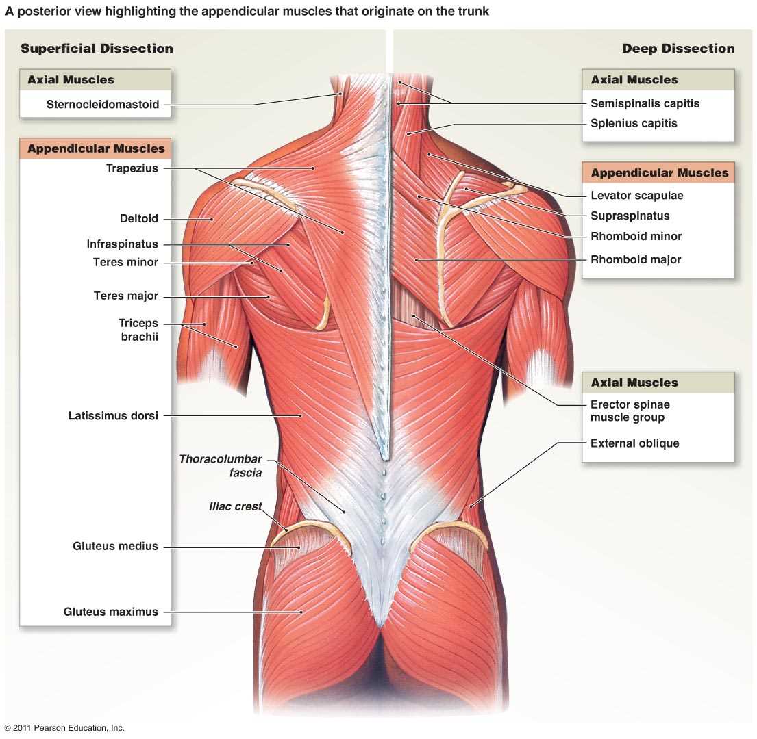
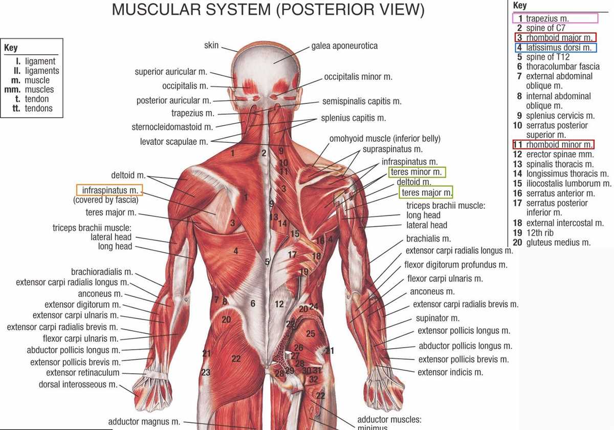
The next part of Exercise 6 focuses on bone markings, which are unique surface features found on bones. Students are required to identify and label various bone markings, such as tuberosities, processes, and foramen. By engaging in this exercise, students develop their anatomical knowledge and further comprehend the roles these bone markings play in muscle attachment, joint formation, and blood vessel or nerve passage.
The final part of Exercise 6 involves the classification of bones, based on their shapes and internal structures. Students are tasked with identifying and classifying different bones into the categories of long bones, short bones, flat bones, and irregular bones. This exercise not only reinforces the knowledge of bone structure but also highlights the functional and structural differences among various bones in the body.
In conclusion, the Human Anatomy and Physiology Lab Manual serves as an invaluable resource for students studying human anatomy and physiology. Exercise 6 supplements their theoretical knowledge with hands-on experience, allowing them to explore bone tissues, bone markings, and the classification of bones. By understanding the answers and explanations of Exercise 6, students can enhance their understanding of the intricate workings of the skeletal system in the human body.
Importance of Human Anatomy and Physiology Lab Manual Exercise 6 Answers
The Human Anatomy and Physiology Lab Manual Exercise 6 focuses on the study of the skeletal system and articulations. This particular exercise allows students to gain hands-on experience and practical knowledge of the various bones, joints, and skeletal structures of the human body. It is an essential component of any anatomy and physiology course as it provides a comprehensive understanding of the structure and function of the skeletal system.
The lab manual provides step-by-step instructions and guidelines for conducting experiments and activities related to the skeletal system. It includes detailed descriptions of each exercise, diagrams, and illustrations to help students visualize and understand the concepts being taught. Additionally, the manual also provides answers to exercises and questions, such as Exercise 6 answers, which are crucial for students to assess their understanding and progress.
The importance of having answers to Exercise 6 in the lab manual cannot be overstated. These answers serve as a valuable resource for students, allowing them to self-assess their performance and identify any knowledge gaps or misconceptions. They provide a benchmark for students to compare their responses with the correct ones, enabling them to gauge their understanding and identify areas that require further study and clarification.
Moreover, having access to Exercise 6 answers helps students reinforce their learning and consolidate their knowledge. By reviewing their answers and comparing them with the correct ones, students can identify their weak points and revise the relevant concepts to improve their understanding. This process allows for active learning and promotes critical thinking skills, as students analyze their mistakes and strive for improvement.
Furthermore, the answers to Exercise 6 in the lab manual equip students with the necessary tools to excel in examinations and assessments. They provide a comprehensive review of the content covered in the exercise, enabling students to revise and reinforce their knowledge before assessments. Understanding the correct answers also helps students understand the expectations of examiners and develop effective strategies for answering similar questions in future assessments.
In conclusion, Exercise 6 answers in the Human Anatomy and Physiology Lab Manual are highly important for students studying the skeletal system. They serve as a self-assessment tool, reinforce learning, and assist students in preparing for examinations. Therefore, it is crucial to utilize the lab manual and its answers to optimize learning outcomes and enhance understanding of human anatomy and physiology.
Understanding Human Anatomy and Physiology
Human anatomy and physiology are crucial topics in understanding the intricate workings of the human body. Anatomy refers to the structure and organization of body parts, while physiology focuses on the functions and processes that occur within those structures. By studying and comprehending human anatomy and physiology, we can gain valuable insights into how our bodies are designed and how they function.
One important aspect of understanding human anatomy and physiology is the ability to identify and label different body parts and systems. This can be achieved through hands-on activities, such as dissection and anatomical models, as well as through the study of diagrams and illustrations. By familiarizing ourselves with the names and locations of various organs, muscles, and bones, we can develop a deeper understanding of the intricate interconnectedness of the human body.
In addition to identifying body parts, it is also essential to comprehend the functions and processes that occur within them. For example, understanding the cardiovascular system allows us to appreciate how the heart pumps blood throughout the body, delivering oxygen and nutrients to our cells. Similarly, knowledge of the respiratory system helps us understand how oxygen is taken in and carbon dioxide is eliminated through the process of breathing.
The Importance of Human Anatomy and Physiology
Studying human anatomy and physiology is not only intriguing, but it also has significant practical implications. Medical professionals, such as doctors and nurses, rely on their understanding of anatomy and physiology to diagnose and treat various health conditions. For example, a doctor needs to know the structure and function of the lungs to diagnose respiratory disorders accurately.
Furthermore, understanding human anatomy and physiology can help us make informed decisions about our own health and well-being. By knowing how our bodies work, we can take proactive steps to maintain good health, such as adopting healthy lifestyle habits and seeking medical care when necessary. A basic knowledge of anatomy and physiology empowers us to be active participants in our own healthcare and make informed choices regarding our bodies.
Role of Lab Manuals in Learning
Lab manuals play a crucial role in enhancing the learning experience in the field of human anatomy and physiology. These manuals serve as a guide for students during their laboratory sessions, providing clear instructions and procedures to perform various experiments and tests. They offer a structured framework that allows students to comprehend and apply theoretical concepts in a practical setting.
One of the key benefits of lab manuals is their ability to improve hands-on learning. By following the step-by-step instructions provided in the manual, students are able to actively engage in the scientific process. They are encouraged to interact with lab equipment, make observations, collect data, and analyze results. This hands-on approach not only deepens their understanding of anatomical structures and physiological processes but also helps in developing critical thinking and problem-solving skills.
Lab manuals also promote collaboration and teamwork among students. Many laboratory exercises require group work, where students work together to complete tasks and achieve common goals. The manual provides a shared reference point for all team members, ensuring that everyone is on the same page and following the same protocols. This collaborative environment fosters effective communication, cooperation, and the sharing of knowledge and ideas.
In addition, lab manuals encourage students to develop a systematic and organized approach to scientific inquiry. The precise instructions, measurements, and recording procedures outlined in the manual help students understand the importance of accuracy and attention to detail in their experiments. It also trains them to follow scientific protocols and adhere to ethical practices in data collection and analysis.
Overview of Exercise 6: The Integumentary System
The integumentary system is one of the most vital systems in the human body, serving as a protective barrier against external threats and regulating body temperature. Exercise 6 focuses on the structure and function of the integumentary system, including the epidermis, dermis, and accessory structures such as hair, nails, and glands.
The exercise begins with a detailed examination of the epidermis, the outermost layer of the skin. Students will learn about the different cell types present in the epidermis, including keratinocytes, melanocytes, and Langerhans cells. They will also explore the layers of the epidermis, understanding the important role of stratum basale in the process of cell renewal.
The dermis, which lies beneath the epidermis, is also an important component of the integumentary system. In this exercise, students will study the various structures found in the dermis, such as blood vessels, nerves, and sensory receptors. They will gain an understanding of the dermis’s role in maintaining skin health and aiding in thermoregulation.
Additionally, this exercise covers the accessory structures of the integumentary system, including hair, nails, and glands. Students will examine the structure and function of hair follicles, learning about the different types of hair and their growth cycles. They will also gain an understanding of the composition and growth of nails, as well as the importance of sweat and oil glands in maintaining skin health.
In summary, Exercise 6 provides a comprehensive overview of the integumentary system, giving students a deeper understanding of the structure and function of the skin and its accessory structures. This knowledge is essential for healthcare professionals and anyone interested in learning about the human body.
Functions and Structure of the Integumentary System
The integumentary system is the largest organ system in the human body, covering and protecting the entire surface area. It consists of the skin, hair, nails, and various glands. This system has multiple functions that are essential for the body’s overall health and well-being.
Protection: One of the primary functions of the integumentary system is to protect the body from external factors such as pathogens, UV radiation, and physical injuries. The skin acts as a barrier, preventing the entry of harmful substances and microorganisms. Additionally, the integumentary system plays a role in regulating body temperature through sweating and the dilation or constriction of blood vessels.
Sensation and Perception: The integumentary system is responsible for the sensory perception of touch, temperature, pressure, and pain. Nerve receptors in the skin transmit these sensations to the brain, allowing us to perceive and interact with the external environment.
Excretion and Secretion: The integumentary system includes various glands that produce and secrete substances necessary for bodily functions. Sweat glands help regulate body temperature by secreting sweat onto the skin’s surface, while sebaceous glands produce and secrete sebum, an oily substance that keeps the skin moisturized and protects it from drying out.
Structure: The integumentary system is composed of three main layers: the epidermis, dermis, and hypodermis. The epidermis is the outermost layer of the skin and is responsible for providing waterproofing and protection. The dermis is located beneath the epidermis and contains blood vessels, nerve endings, and hair follicles. The hypodermis, also known as the subcutaneous tissue, is the deepest layer and consists of fat cells that provide insulation and serve as an energy reserve.
In conclusion, the integumentary system plays a crucial role in the overall functioning of the body. It not only protects us from external threats but also allows us to sense and interact with the world around us. Understanding the functions and structure of this system is essential for maintaining our health and well-being.
The Importance of Exercise 6 in Understanding the Integumentary System
The integumentary system is composed of the skin, hair, nails, and various glands. It plays a vital role in protecting the body from external factors such as pathogens, UV radiation, and physical damage. In order to fully understand the integumentary system and its functions, it is important to engage in practical exercises like Exercise 6 in the Human Anatomy and Physiology lab manual.
Exercise 6 focuses on the structure and function of the skin, which is the largest organ of the human body and a major component of the integumentary system. By conducting the exercises outlined in this exercise, students can gain a hands-on and visual understanding of the different layers of the skin, the structures within each layer, and their respective functions.
- Observing the Epidermis: Through exercises like the use of a microscope to examine skin slides, students can observe the different layers of the epidermis, including the stratum basale, stratum spinosum, stratum granulosum, and stratum corneum. This allows them to understand the process of keratinization and appreciate the role of each layer in providing protection and waterproofing.
- Assessing Skin Variations: Exercise 6 also introduces students to variations in skin color, texture, and markings. By examining samples and noting differences in pigmentation, hair distribution, and the presence of freckles or birthmarks, students can gain an understanding of the influence of genetics and environmental factors on the integumentary system.
- Exploring Accessory Structures: The integumentary system includes various accessory structures such as hair follicles, sweat glands, and sebaceous glands. Exercise 6 provides students with the opportunity to examine these structures under a microscope, allowing them to understand their roles in regulating body temperature, lubricating the skin, and protecting against pathogens.
Overall, Exercise 6 in the Human Anatomy and Physiology lab manual is essential in deepening students’ understanding of the integumentary system. By actively engaging in hands-on activities and visualizing the structures and functions of the skin, students can develop a comprehensive knowledge of this complex system and its importance in maintaining homeostasis and protecting the body.