
Preparing for an Anatomy and Physiology 1 Exam 2 can be a daunting task, but with the right study strategies and a solid understanding of the material, success can be within reach. This comprehensive guide aims to provide students with the essential information and resources needed to excel in this challenging exam.
Exam 2 in Anatomy and Physiology 1 typically covers a wide range of topics, including the muscular system, the nervous system, and sensory organs. Understanding the intricate details of muscle structure and function, the pathways and mechanisms of the nervous system, and the complexities of the sensory organs is crucial for success in this exam.
To effectively prepare for this exam, it is important to utilize a variety of study techniques. This may include creating colorful and detailed diagrams that illustrate the different muscle groups and their functions, reviewing lecture notes and textbooks, engaging in group discussions to reinforce understanding, and utilizing online resources, such as video tutorials and interactive quizzes, to test knowledge and identify areas that require further review.
Furthermore, practicing with past exam questions and participating in mock exams can help familiarize students with the format and structure of the actual exam, allowing them to develop time-management skills and assess their level of preparedness. Additionally, seeking assistance from professors or teaching assistants for clarification on difficult concepts can be immensely beneficial.
Anatomy and Physiology 1 Exam 2
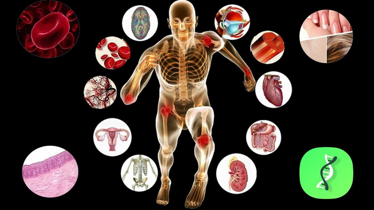
Anatomy and Physiology 1 Exam 2 is a comprehensive examination that assesses students’ understanding of the anatomical structures and physiological processes of the human body. This exam covers various body systems, including the cardiovascular, respiratory, digestive, and urinary systems, as well as the integumentary and musculoskeletal systems.
Students are tested on their knowledge of the anatomy and functions of organs, tissues, and cells within these systems, as well as their ability to apply this knowledge to real-world scenarios. They are also required to demonstrate their understanding of various physiological processes, such as the mechanisms of respiration, digestion, and blood circulation.
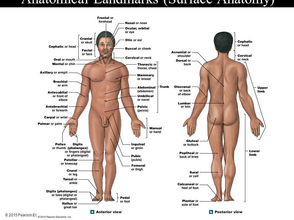
The exam typically consists of a combination of multiple-choice, short answer, and essay questions. Students are expected to have a strong foundation in the basic principles of anatomy and physiology, including terminology, anatomical position, and the structure and function of cells and tissues. They should also be able to interpret anatomical diagrams, such as cross-sectional views of organs and body systems.
- Topics covered in Anatomy and Physiology 1 Exam 2 may include:
- Cardiovascular system: structure and function of the heart, blood vessels, and blood circulation.
- Respiratory system: anatomy and physiology of the lungs, breathing mechanisms, and gas exchange.
- Digestive system: structure and function of the digestive organs, nutrient absorption, and digestion processes.
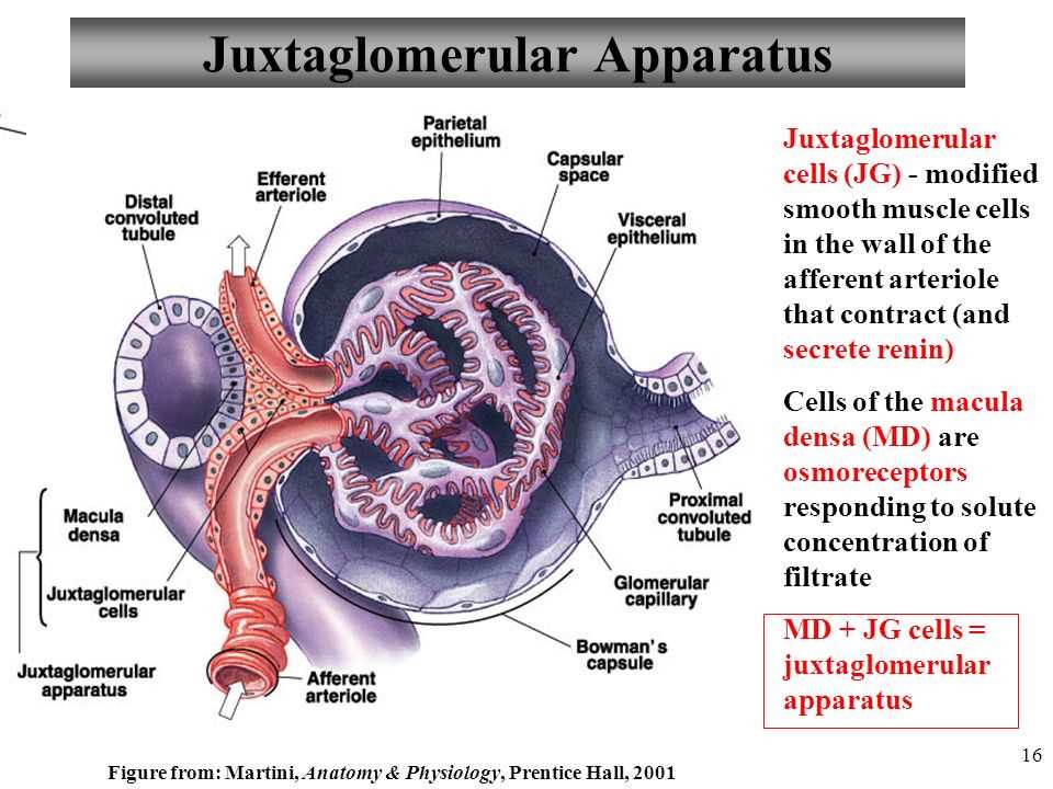
- Urinary system: anatomy and physiology of the kidneys, urine formation, and regulation of fluid balance.
- Integumentary system: structure and function of the skin, hair, and nails.
- Musculoskeletal system: anatomy and physiology of the muscles, bones, and joints.
Preparing for Anatomy and Physiology 1 Exam 2 requires active studying, which may include reviewing lecture notes, completing textbook readings, and practicing with sample questions. It is important for students to understand not only the structure and function of each body system but also their interrelationships and how they contribute to overall body homeostasis.
By mastering the content covered in Anatomy and Physiology 1 Exam 2, students will be well-prepared to continue their studies in advanced anatomy and physiology courses or pursue careers in healthcare, such as nursing, medicine, or physical therapy.
The Structure and Function of the Cell
The cell is the basic structural and functional unit of all living organisms. It is a highly organized and complex structure that carries out all the activities necessary for life. The understanding of the cell’s structure and function is crucial in the field of anatomy and physiology.
The cell is enclosed by a plasma membrane, which separates the cell from its external environment and controls the passage of substances in and out of the cell. It is composed of a phospholipid bilayer with embedded proteins that allow for the transport of molecules and ion channels. The plasma membrane also contains receptor proteins that allow the cell to respond to signals from its environment.
Cell organelles and their functions:
- Nucleus: The nucleus houses the cell’s genetic material, DNA, and is responsible for controlling the activities of the cell. It is surrounded by a nuclear envelope and contains the nucleolus, where ribosomes are synthesized.
- Mitochondria: Mitochondria are the powerhouses of the cell, as they are responsible for producing energy in the form of ATP through cellular respiration. They have a double membrane and contain their own DNA.
- Endoplasmic reticulum: The endoplasmic reticulum is a network of membranes that is involved in the synthesis, modification, and transport of proteins and lipids. It can be rough, with ribosomes attached, or smooth, without ribosomes.
- Golgi apparatus: The Golgi apparatus is responsible for modifying, sorting, and packaging proteins and lipids for transport within the cell or secretion outside the cell.
- Lysosomes: Lysosomes contain digestive enzymes and are involved in the breakdown of waste materials, cellular debris, and pathogens. They also play a role in cell death and recycling of cellular components.
- Cytoskeleton: The cytoskeleton provides structural support and shape to the cell. It is composed of microtubules, microfilaments, and intermediate filaments, which also help with cell movement and intracellular transport.
These are just a few examples of the many organelles present in a cell. Each organelle has specific functions that contribute to the overall structure and function of the cell, allowing it to carry out its various activities and maintain homeostasis.
Tissues: Types and Characteristics
Tissues are groups of cells with similar structure and function that work together to perform specific tasks in the body. There are four main types of tissues: epithelial, connective, muscle, and nervous. Each type of tissue has its own unique characteristics and functions.
Epithelial tissue covers the surfaces of the body and lines the organs and cavities. It has tightly packed cells that form a protective barrier. Epithelial tissue can be classified into different types based on its shape and arrangement of cells, such as squamous, cuboidal, and columnar. This type of tissue is important for functions such as absorption, secretion, and protection.
Connective tissue provides support and connects different structures in the body. It consists of cells and extracellular matrix, which is made up of proteins and fibers. Connective tissue can be classified into various types, including loose connective tissue, dense connective tissue, and specialized connective tissues like cartilage, bone, and blood. Connective tissue plays a role in functions such as structural support, insulation, and transportation of nutrients.
… continue reading on the topic of muscle and nervous tissue …
The Skeletal System: Bones, Joints, and Movement
The skeletal system is composed of bones, joints, and connective tissues that provide support, protection, and movement for the body. The bones in the skeletal system provide a framework for the body, allowing it to maintain its shape and structure. They also protect vital organs, such as the brain and heart, from injury. Additionally, the bones store minerals, such as calcium and phosphorus, which are essential for maintaining overall health and function.
There are different types of bones in the skeletal system, including long bones, short bones, flat bones, and irregular bones. Long bones, such as the femur and humerus, are responsible for supporting weight and facilitating movement. Short bones, such as those found in the wrists and ankles, provide stability and support for the body. Flat bones, such as the skull and ribs, protect vital organs and provide surfaces for muscle attachment. Irregular bones, such as the vertebrae and pelvic bones, have unique shapes and functions within the skeletal system.
Joints are the points where two or more bones meet and allow for movement. There are different types of joints in the skeletal system, including immovable joints, slightly movable joints, and freely movable joints. Immovable joints, also known as synarthroses, do not allow for any movement, such as the joints between the bones of the skull. Slightly movable joints, also known as amphiarthroses, allow for limited movement, such as the joints between the vertebrae. Freely movable joints, also known as diarthroses, allow for a wide range of movement and are found in the limbs, such as the shoulder and hip joints.
In order for movement to occur, muscles must be able to contract and pull on the bones. This is made possible by the attachment of muscles to bones through tendons. Tendons are tough connective tissues that connect muscles to bones and allow for movement. When a muscle contracts, it pulls on the tendon, which in turn pulls on the bone, causing movement. This coordinated effort between muscles, tendons, and bones allows for the wide range of movements and activities that the human body is capable of.
The Muscular System: Types of Muscles and Muscle Contraction
The muscular system is responsible for producing movement, maintaining posture, and generating heat in the body. There are three types of muscles in the body: skeletal muscles, smooth muscles, and cardiac muscles. Each type of muscle has its own unique structure and function.
Skeletal muscles are attached to bones and are responsible for voluntary movements. They are made up of long, cylindrical cells called muscle fibers. These muscles have a striated appearance due to the repeating pattern of actin and myosin filaments. Skeletal muscles are controlled by the somatic nervous system and are under conscious control.
Smooth muscles, also known as involuntary muscles, are found in the walls of organs and blood vessels. They have a non-striated appearance and are controlled by the autonomic nervous system. Smooth muscles function involuntarily to control processes such as digestion, blood flow, and breathing.
Cardiac muscles are found exclusively in the heart and are responsible for the involuntary contraction of the heart. They have a striated appearance and are controlled by the autonomic nervous system. Cardiac muscles are unique in that they have intercalated discs, specialized structures that allow for rapid communication between individual muscle cells.
Muscle contraction is a complex process that involves the interaction of actin and myosin filaments. When a muscle is stimulated by a nerve impulse, calcium ions are released from the sarcoplasmic reticulum, a specialized organelle within muscle cells. The calcium ions bind to a protein called troponin, which allows myosin filaments to attach to actin filaments. As the myosin filaments slide past the actin filaments, the muscle contracts and generates force.
- Skeletal muscles: attached to bones, voluntary control
- Smooth muscles: in organs and blood vessels, involuntary control
- Cardiac muscles: in the heart, involuntary control
In summary, the muscular system consists of three types of muscles: skeletal, smooth, and cardiac. Each type of muscle has its own structure and function. Muscle contraction is a complex process that involves the interaction of actin and myosin filaments. Understanding the different types of muscles and how they contract is essential for comprehending the functioning of the muscular system.
The Nervous System: Neurons, Synapses, and Nerve Impulses
The nervous system is a complex network of specialized cells called neurons that transmit signals throughout the body. Neurons have a unique structure consisting of a cell body, dendrites, and an axon. The cell body contains the nucleus and other organelles necessary for the neuron’s functions. Dendrites receive signals from other neurons, while the axon carries signals away from the cell body.
Neurons communicate with each other at specialized junctions called synapses. At a synapse, the axon terminal of one neuron releases neurotransmitters, which are chemical messengers that cross the synapse and bind to receptors on the dendrites of another neuron. This transfer of information allows signals to be transmitted from one neuron to another.
In order for a signal to travel along a neuron, a nerve impulse is generated. This impulse is an electrical charge that travels down the axon of the neuron. The generation of a nerve impulse relies on a difference in electrical charge between the inside and outside of the neuron, known as the resting membrane potential. When a neuron receives a signal, its membrane becomes permeable to certain ions, causing an electrical imbalance and the creation of an action potential, or nerve impulse.
Overall, the nervous system plays a crucial role in controlling and coordinating the body’s activities. Neurons, synapses, and nerve impulses are essential components of this system, allowing for communication between different parts of the body and facilitating the transmission of important signals.
The Endocrine System: Hormones and their Effects
The endocrine system is a complex network of glands and organs that produce and release hormones into the bloodstream. These hormones act as chemical messengers, traveling through the body to target cells and organs, where they regulate various physiological processes.
Hormones are substances that are produced in one part of the body and have effects in other parts of the body. They are typically released in response to specific stimuli, such as stress, low blood sugar, or changes in light and temperature. Hormones can have a wide range of effects, including regulating metabolism, growth and development, sexual function, and mood.
Examples of hormones and their effects:
- Insulin: produced by the pancreas, insulin helps regulate blood sugar levels by promoting the uptake of glucose from the blood into cells.
- Thyroid hormones: produced by the thyroid gland, these hormones regulate metabolism, energy production, and growth and development.
- Estrogen and progesterone: produced by the ovaries, these hormones play a key role in regulating the menstrual cycle and maintaining pregnancy.
- Cortisol: produced by the adrenal glands, cortisol helps the body respond to stress by increasing blood sugar levels and suppressing the immune system.
Hormones can have both short-term and long-term effects on the body. Some effects may be immediate, such as an increase in heart rate or a surge of energy, while others may take longer to manifest, such as changes in bone density or muscle mass. Hormones also interact with each other, influencing their effects and creating a complex web of hormonal regulation.